Definition of "Pulse"
Pulse is measuring heart beat by palpating a peripheral artery by the fingertip (with the exception of using the thumb). Sometimes, there is delay, which is indicative of pathology.
Pulses can be palpated at any place that allows an artery to be compressed against a bone, including:
Head and neck:
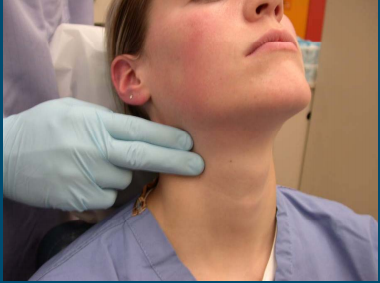
- Carotid artery, located in the neck, between the anterior border of the sternocleidomastoid muscle, above the hyoid bone, and laterla to the thyroid cartilage. It should be palpated gently while the patient is sitting or lying down. Stimulating its baroreceptors with low palpation can provoke severe bradycardia, or even stop the heart in sensitive patients. A patient's 2 carotid arteries should NOT be palpated at the same time, as it may limit blood flow to the head, possibly causing fainting or brain ischemia
Source: ClassConnection
- Facial artery, located on the mandible (lower jawbone), on a line with the corners of the mouth
- [Superficial] temporal artery, located on the temple directly in front of the ear
Upper limb:
- Axillary pulse, located inferiorly of the lateral wall of the axilla
- Brachial artery, located on the inside of the upper arm, inside the elbow, frequently used in place of carotid pulse in infants
Source: ClassConnection
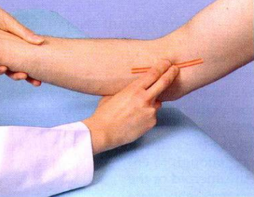
- Radial artery, located on the lateral of the wrist, at the anatomical snuffbox, commonly measured using 3 fingers, so the finger closest to the heart occludes the pulse pressure, the middle finger otains a crude estimate of blood pressure, and the ring finger is used to nullify the effect of the ulnar pulses as the 2 arteries are connected via the palmar arches
- Ulnar artery, located on the medial of the wrist

Source: 2013/03/Radial-pulse.jpg">EasyMBBS
Torso:
- Apical pulse, located at the 5th intercostal space, 1.25cm lateral to the midclavicular line. Unlike other pulse sites, it is not under an artery, but at the apex of the heart
Lower limb:
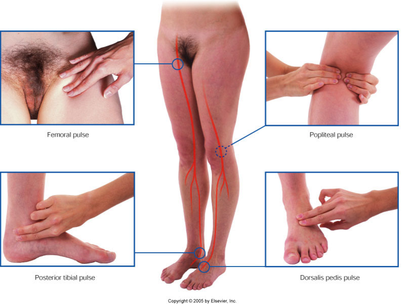
- Femoral artery, located at the groin, in the inner thigh, at the mid-inguinal point, halfway between the pubic symphysis and ASIS (anterior superior iliac spine)
- Popliteal artery, located above and behind the knee, in the popliteal fossa, found by holding the bent knee. The knee is bent at apprximately 124 degres, and the doctor holds it in both ahdns to find the popliteal artery in the pit behind th eknee

Source: GLA
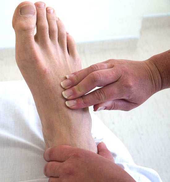
- Dorsalis pedis artery (on the foot), is located on top of the foot, immediately lateral to the extensor of hallucis longus
Source: OSCE skills
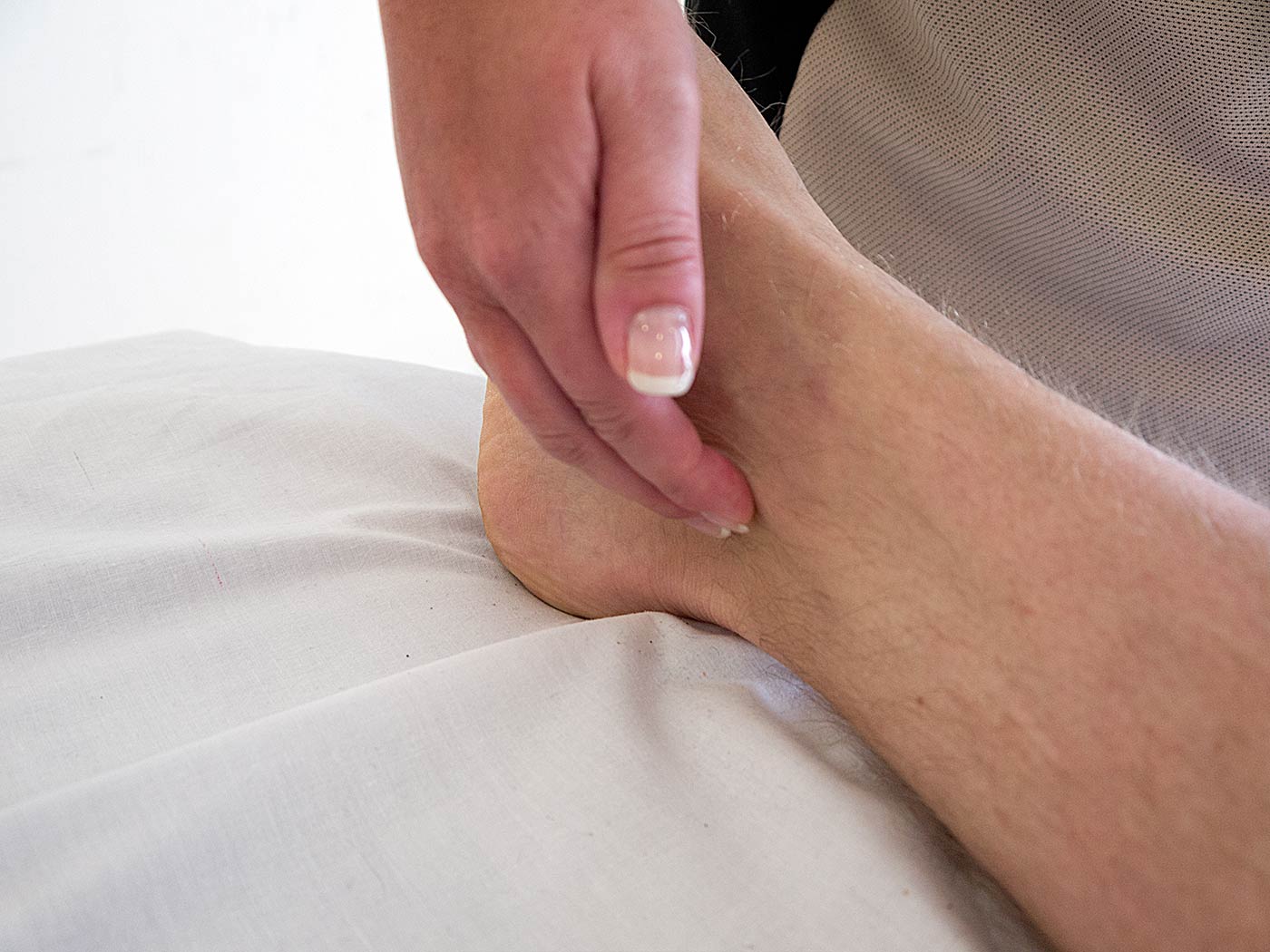
- Posterior tibial artery, located on the medial side of the ankle joint, over Pimenta's Point, where 3 fingers are placed at the midpoint of an imaginary line drawn between the bony prominence of the medial maleolus, and the insertion of the achilles tendon
Source: GLA
Source: Elsevier
HR can also be measured by auscultating the heart beat using a stethoscope.
Patient information
What is pulse? It's the beating of the heart that you're feeling, right?Yep. So you're feeling the heart beat peripherally.
How do you feel the heart beating peripherally?
So you press the artery against a bone. You can do this in the head, at the carotid artery in the neck. You can do this in the arm, at the brachial artery inside the elbow, radial artery at the wrist. You can also do this in the feet, at the femoral artery at the groin, popliteal artery behind the knee, posterior tibial artery near the ankle joint, and dorsalis pedis artery on the foot.
- Rate, is in beats per minute (bpm), representing heart rate. It has the extremities of bradycardia and tachycardia
- Rhythm can either be:
- Regular
- Regularly irregular, is a regular but intermittent pulse, and can be caused by:
- Pulsus bigeminus
- 2nd degree AV block
- Irregularly irregular, which is irregularly and intermittent pulse, can be caused by:
- Atrial fibrillation
- Volume (aka amplitude, expansion, size of pulse), is the degree of expansion of the artery during diastole and systole. It includes:
- Hypokinetic pulse (aka weak pulse), indicates narrow pulse pressure. It can be caused by:
- Low cardiac output, e.g. shock, CHF
- Hypovolemia
- Valvular heart disease, e.g. aortic outflow tract obstruction, mitral stenosis, aortic arch syndrome
- Hyperkinetic pulse (aka bounding pulse), indicates high pulse pressure. It can be caused by:
- Low peripheral resistance, e.g. fever, anemia, thyrotoxicosis, AV fistula, Paget's disease, beriberi, liver cirrhosis
- Increased cardiac output
- Increased stroke volume, e.g. anxiety, exercise, complete heart block, aortic regurgitation
- Decreased distensibility of arterial system, e.g. atherosclerosis, HTN, and coarctation of aorta
- Hypokinetic pulse (aka weak pulse), indicates narrow pulse pressure. It can be caused by:
- Force (aka compressibility of pulse), is a rough measure of systolic BP
- Tension, coresponds to diastolic BP. It includes:
- Pulsus mollis (low tension pulse), where the vessel is soft or impalpable between beats
- Pulsus durus (high tension pulse), where the vessels feel rigid even between pulse beats
- Equality/delay, comparing pulses at different places
- A discrepant/unequal pulse between the L and R radial artery, indicates:
- Anomalous/aberrant course of artery
- Coarctation of aorta
- Aortitis
- Dissecting aneurysm
- Peripheral embolism
- Unequal pulse between upper and lower extremities, e.g. radio-femoral delay, is seen in:
- Coarctation of aorta, where the femoral pulse may be significantly delayed compared to the radial pulse
- Supravalvar aortic stenosis
- Aortitis
- Block at bifurcation of aorta
- Dissection of aorta
- Iatrogenic trauma
- Arteriosclerotic obstruction
- A discrepant/unequal pulse between the L and R radial artery, indicates:
- Compressibility, as a normal artery is not palpable after flattening by digital pressure. A thick radial artery palpable 7.5-10cm up the forearm is suggestive of arteriosclerosis
- Doppler auscultation
- Tachycardia (higher than normal)
Find a practitioner
Practitioner count: 0